


The Carl Zeiss photography competition at the University of Cambridge announced its results last month. The annual competition, meant for only staff and students at the Department of Engineering, claims to showcase in images the scope of engineering research.
The 11-year-old competition is not just about photography. At a nano-level, photo manipulation and digital art become necessary to visualise engineering concepts in an appealing – and sometimes new – manner. The competition is sponsored by German opticals manufacturer Carl Zeiss and is judged within the university.
Here is a selection of entries (and the winners), from the competition's Flickr page.
This is a false colored low magnification electron micrograph of free standing graphene foam. Graphene foam is made by growing a few layers of graphene on the surface of a porous metal foam skeleton using chemical vapour deposition technique. The metal foam skeleton is then removed by carefully dissolving it in an etching solution. Because of its unique properties, e.g. electrically conductive, highly porous, and lightweight, graphene foam has the potential to be used in numerous advanced applications including chemical sensing, energy storage, and ultra-lightweight structures. Submitted by Adrianus Indrat Aria.
New techniques in machine learning and image processing allow us to extrapolate the scene of a painting to see what the full scenery might have looked like. We used the PatchMatch algorithm on the frame of van Gogh's painting "Starry Night" to extrapolate its contents. The second image (II) is the same piece with a "frame" added using photoshop to distinguish the painting from the extrapolated background. Submitted by Yarin Gal.
This SEM image shows formation of a crack bridging for self-healing in concrete. These were taken from the self-healed samples to investigate the self-healing materials formation in the concrete cracked zone. The research is aimed to investigate the improvement of autogenous self-healing capability of Ordinary Portland cement adding optimum amount of reactive Magnesia (MgO) based materials. Submitted by Tanvir Qureshi.
An array of diffractive lenses imaged using dark field optical microscopy. Each lens is composed of hundreds of sub-wavelength metallic nanostructures which scatter light at specific wavelengths. The lens pattern is designed such that the light experiences a different phase profile across this planar structure resulting in constructive inference forming a bright focal point. This interesting image represents a failed fabrication attempt in which the photoresist used in the lithography process has not worked correctly. Submitted by Calum Williams, Yunuen Montelongo and Jaime Tenorio-Pearl.
The Royal Mail Tunnel site: A site that has been a showcase for our technology and monitoring innovation. I have worked with two other PhD students on this project to try and capture a comprehensive catalogue of very discrete movements in order to determine how the tunnel is actually behaving, which is unknown at this level of detail. Submitted by Matthew Wilcock.
This image shows the interesting optical effects produced by shining a typical red laser pointer (at top-right) through a standard set of digital camera lenses. The grid of spots surrounding the laser light at top-right is the diffraction from the surface of the camera sensor due to the focused laser light that is then reflected off the sensor's covering glass and re-imaged by the sensor. The round ghostly patterns in the middle are the various lens elements made visible due to phase differences of the lenses interfering with each other. Focused laser speckle is also visible at the bottom-left as well as lens flare from light scattered in the lens elements. Submitted by Ananta Palani.
Nanomaterials such as carbon nanotubes and graphene, are not only interesting from a scientific or industrial perspective, they can sometimes also form beautiful structures, which under electron microscopes reveal glimpses of a fascinating nanoworld. The structures on display are each about one thousandth of a millimeter large, and consist of thousands of nanoparticles. Submitted by Michael De Volder.
In this picture the invisible has been made visible. A web of carbon nanotubes that bundled and are integrated in a matrix is formed. The carbon nanotubes were synthesized in a continuous gas phase process and the picture shown is a snapshot of this. The picture is taken from a part of the worktube and its actual height is 50 mm. The image is a negative. Submitted by Christian Hoecker.
We welcome your comments at
letters@scroll.in.
The 11-year-old competition is not just about photography. At a nano-level, photo manipulation and digital art become necessary to visualise engineering concepts in an appealing – and sometimes new – manner. The competition is sponsored by German opticals manufacturer Carl Zeiss and is judged within the university.
Here is a selection of entries (and the winners), from the competition's Flickr page.

Adrianus Indrat Aria: Asteroidea Electrica, First Prize in the ZEISS photography competition 2014.
This is a false colored low magnification electron micrograph of free standing graphene foam. Graphene foam is made by growing a few layers of graphene on the surface of a porous metal foam skeleton using chemical vapour deposition technique. The metal foam skeleton is then removed by carefully dissolving it in an etching solution. Because of its unique properties, e.g. electrically conductive, highly porous, and lightweight, graphene foam has the potential to be used in numerous advanced applications including chemical sensing, energy storage, and ultra-lightweight structures. Submitted by Adrianus Indrat Aria.

Yarin Gal: Extrapolated art II, Second prize in the ZEISS photography competition 2014.
New techniques in machine learning and image processing allow us to extrapolate the scene of a painting to see what the full scenery might have looked like. We used the PatchMatch algorithm on the frame of van Gogh's painting "Starry Night" to extrapolate its contents. The second image (II) is the same piece with a "frame" added using photoshop to distinguish the painting from the extrapolated background. Submitted by Yarin Gal.

Tanvir Qureshi: Concrete Crack Bridge for Self-Healing I
This SEM image shows formation of a crack bridging for self-healing in concrete. These were taken from the self-healed samples to investigate the self-healing materials formation in the concrete cracked zone. The research is aimed to investigate the improvement of autogenous self-healing capability of Ordinary Portland cement adding optimum amount of reactive Magnesia (MgO) based materials. Submitted by Tanvir Qureshi.

Calum Williams, Yunuen Montelongo & Jaime Tenorio-Pearl: A world of ruined lenses II.
An array of diffractive lenses imaged using dark field optical microscopy. Each lens is composed of hundreds of sub-wavelength metallic nanostructures which scatter light at specific wavelengths. The lens pattern is designed such that the light experiences a different phase profile across this planar structure resulting in constructive inference forming a bright focal point. This interesting image represents a failed fabrication attempt in which the photoresist used in the lithography process has not worked correctly. Submitted by Calum Williams, Yunuen Montelongo and Jaime Tenorio-Pearl.

Matthew Wilcock: Royal Mail Tunnel
The Royal Mail Tunnel site: A site that has been a showcase for our technology and monitoring innovation. I have worked with two other PhD students on this project to try and capture a comprehensive catalogue of very discrete movements in order to determine how the tunnel is actually behaving, which is unknown at this level of detail. Submitted by Matthew Wilcock.

Ananta Palani: Invisible Made Visible III
This image shows the interesting optical effects produced by shining a typical red laser pointer (at top-right) through a standard set of digital camera lenses. The grid of spots surrounding the laser light at top-right is the diffraction from the surface of the camera sensor due to the focused laser light that is then reflected off the sensor's covering glass and re-imaged by the sensor. The round ghostly patterns in the middle are the various lens elements made visible due to phase differences of the lenses interfering with each other. Focused laser speckle is also visible at the bottom-left as well as lens flare from light scattered in the lens elements. Submitted by Ananta Palani.
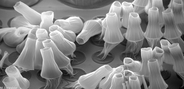
Michael De Volder: Carbon Nanotube Lanterns
Nanomaterials such as carbon nanotubes and graphene, are not only interesting from a scientific or industrial perspective, they can sometimes also form beautiful structures, which under electron microscopes reveal glimpses of a fascinating nanoworld. The structures on display are each about one thousandth of a millimeter large, and consist of thousands of nanoparticles. Submitted by Michael De Volder.

Christian Hoecker: Web of Science II
In this picture the invisible has been made visible. A web of carbon nanotubes that bundled and are integrated in a matrix is formed. The carbon nanotubes were synthesized in a continuous gas phase process and the picture shown is a snapshot of this. The picture is taken from a part of the worktube and its actual height is 50 mm. The image is a negative. Submitted by Christian Hoecker.